

Peptides vs Proteins: Key Differences Explained
Peptides are short chains of amino acids, while proteins are longer chains that fold into complex, functional macromolecules. The standard shorthand places the boundary at roughly 50 amino acids; molecular weight thresholds vary by source without a fixed cutoff. That cutoff is a working convention, not a hard chemical law, and you will find different thresholds across textbooks and research fields.
The practical distinctions, at a glance:
- Size: Peptides are typically composed of 2–50 amino acid residues; proteins are generally 50 or more residues in length, though some sources set the protein threshold as high as 100 residues.
- Structure: Peptides rarely fold into stable three-dimensional domains; proteins adopt secondary, tertiary, and sometimes quaternary structures that enable complex functions.
- Function: Peptides act mainly as signals and modulators; proteins serve as enzymes, structural scaffolds, transporters, and antibodies.
- Stability: Short peptides are more susceptible to enzymatic degradation than most folded proteins.
- Production: Peptides are commonly synthesized chemically; proteins are usually produced by recombinant expression in living cells.
Table of Contents
- What are peptides and proteins, and how do their sizes differ?
- How does structural complexity separate peptides from proteins?
- What biological roles do peptides and proteins each perform?
- How are peptides and proteins made in the body and in the lab?
- How do scientists distinguish peptides from proteins in the lab?
- Common examples and misconceptions about peptides and proteins
- When should you call a chain a peptide versus a protein?
- Why peptide purity and analytical verification matter for research
- Key Takeaways
- Why the peptide-protein distinction still matters
- Useful sources for further reading
What are peptides and proteins, and how do their sizes differ?
The genome.gov genetics glossary defines a peptide as a short chain of amino acids linked by peptide bonds. A protein, by the same authority, is a longer chain that folds into a functional macromolecule capable of performing diverse cellular roles.
The most widely cited numeric threshold puts peptides at 2–50 residues and proteins at 50 or more. Some authors shift that boundary to 40 or even 100 residues depending on the research context. Molecular weight offers a parallel frame: molecules below roughly 5,000 Da are often called peptides, while those above that mark are proteins. ScienceDirect’s overview of peptides and proteins notes that insulin, frequently cited as the smallest true protein, weighs approximately 5,800 Da, which places it right at the boundary.
The intermediate term polypeptide covers chains that are too long to be called a simple peptide but may not yet fold into a stable protein structure. Oligopeptide refers to very short chains of just a few residues. Britannica’s explainer on the peptide-protein distinction is direct on this point: these categories are functional classifications, not strict chemical divisions. Context matters, and a molecule described as a “peptide” in a pharmacology paper might be labeled a “small protein” in a structural biology journal.
How does structural complexity separate peptides from proteins?
Biochemists describe protein architecture across four levels. Primary structure is simply the sequence of amino acids. Secondary structure refers to local folding patterns, mainly alpha-helices and beta-sheets, stabilized by hydrogen bonds. Tertiary structure is the full three-dimensional shape of a single polypeptide chain. Quaternary structure arises when two or more folded chains assemble into a larger complex.
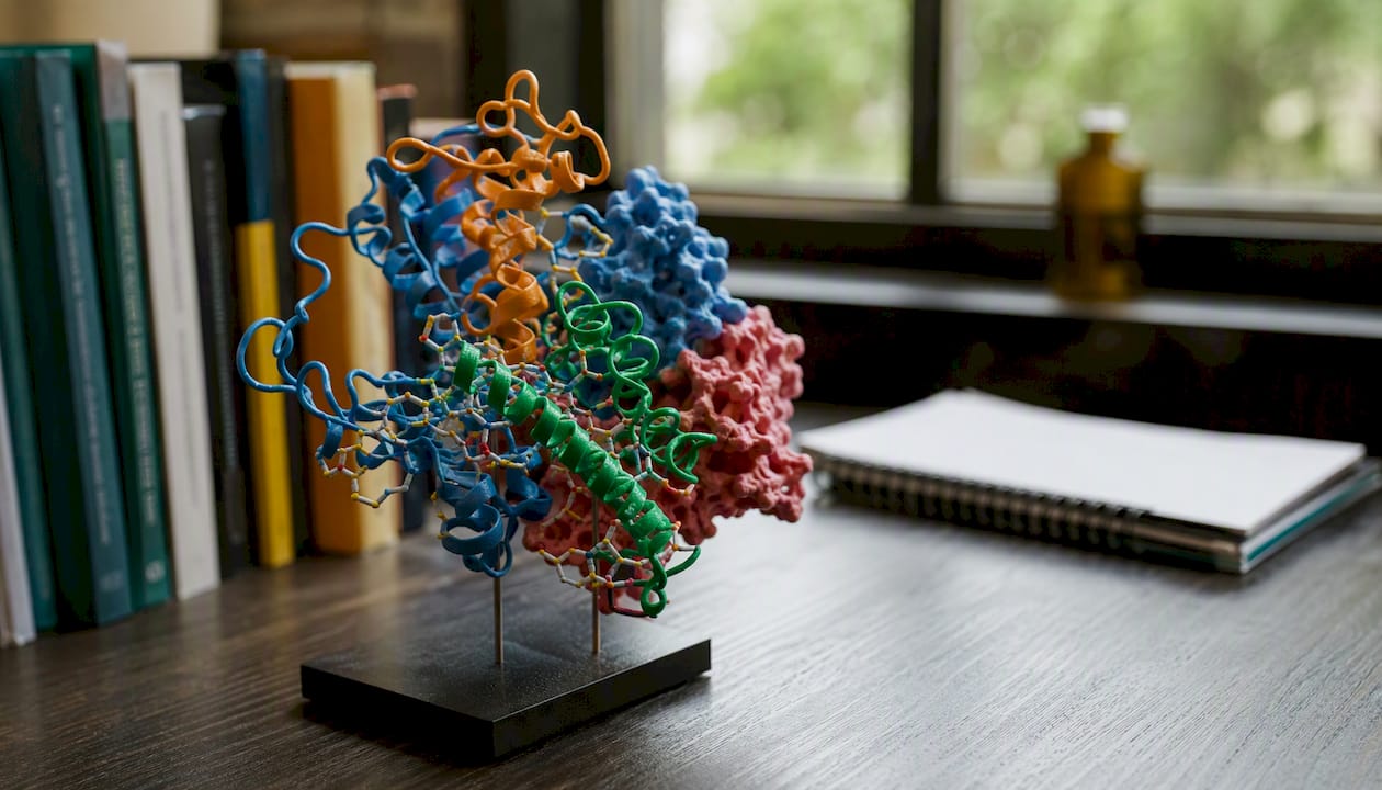
Short peptides typically express only primary structure and, occasionally, limited secondary structure. They rarely achieve stable tertiary folding because the chain is too short to form the hydrophobic core that anchors a protein’s three-dimensional shape. NCBI Bookshelf’s chapter on protein structure and function explains that this folding hierarchy is what allows proteins to build precise active sites and structural domains, capabilities that short peptides simply cannot replicate.
Consider the contrast between a serine protease enzyme and the nine-residue signaling peptide oxytocin. The enzyme folds into a stable tertiary structure with a catalytic triad, a geometry that positions three specific amino acids within angstroms of each other to cleave peptide bonds with high specificity. Oxytocin, by contrast, acts by binding a receptor at the cell surface. Its function depends on sequence and shape, but not on the kind of stable internal architecture that makes enzymatic catalysis possible.
What biological roles do peptides and proteins each perform?
Functional differences follow directly from structural ones. Peptides tend to act as fast, targeted signals; proteins take on the heavy structural and catalytic work of the cell.
Common peptide functions:
- Signaling hormones (insulin, glucagon, GLP-1 analogs)
- Neuropeptides and neurotransmitter modulators (oxytocin, substance P)
- Antimicrobial peptides that disrupt bacterial membranes (defensins, magainins)
- Growth factors and short-range cell modulators (BPC-157, TB-500 in research contexts)
Common protein functions:
- Enzymes that catalyze metabolic reactions (DNA polymerase, amylase)
- Structural components (collagen in connective tissue, keratin in hair and nails)
- Oxygen transport (hemoglobin carries O₂ through the bloodstream)
- Immune defense (antibodies, complement proteins)
- Molecular transport and signaling receptors
Michigan State University’s chemistry resource on proteins illustrates this breadth well: collagen, the most abundant protein in the human body, provides tensile strength to tendons and skin through a triple-helix quaternary structure, while hemoglobin’s four-subunit assembly allows cooperative oxygen binding across a range of partial pressures. Neither function would be achievable by a short peptide chain.
How are peptides and proteins made in the body and in the lab?
Both peptides and proteins originate from the same cellular machinery. Ribosomes translate messenger RNA into a linear amino acid sequence, producing a polypeptide chain. For many signaling peptides, the ribosome first builds a larger precursor, called a preproprotein or propeptide, which is then cleaved by proteases to release the active, shorter peptide. Insulin follows exactly this path: the ribosome produces preproinsulin, which is processed to proinsulin and then to mature insulin by removal of the C-peptide.
Post-translational modifications add another layer of complexity. Glycosylation attaches sugar chains, phosphorylation adds phosphate groups, and disulfide bond formation locks certain peptides and proteins into stable conformations. These modifications are often critical for biological activity and are difficult to replicate outside a living cell.
In the laboratory, the production route diverges sharply by size. NCBI Bookshelf’s chapter on peptide synthesis and purification describes solid-phase peptide synthesis (SPPS) as the standard method for producing peptides chemically. SPPS builds the chain one residue at a time on a solid resin support, cycling through deprotection, activation, and coupling steps. The method is practical for chains up to roughly 50 residues. Longer proteins, however, require recombinant expression in bacterial, yeast, or mammalian cell systems because chemical synthesis becomes inefficient and error-prone at that scale.
How do scientists distinguish peptides from proteins in the lab?
| Method | What it measures | Best used for |
|---|---|---|
| Mass spectrometry (MS) | Exact molecular mass and sequence fragments | Confirming identity and molecular weight; distinguishing peptides from proteins at the boundary |
| HPLC | Purity by retention time; separation of components | Quantifying purity (% area), detecting impurities, verifying batch consistency |
| SDS-PAGE | Apparent molecular weight by gel migration | Rapid size estimation; less accurate below ~5 kDa |
| Size-exclusion chromatography (SEC) | Hydrodynamic radius / molecular size in solution | Detecting aggregation; separating peptides from larger protein contaminants |
| Circular dichroism (CD) | Secondary structure content (helix, sheet, random coil) | Confirming folding state; useful for proteins and structured peptides |
Mass spectrometry and HPLC are the primary tools for confirming whether a molecule is a peptide or a small protein, and molecular weight from MS is the most direct evidence. The University of Queensland’s IMB explainer on peptides vs proteins notes that analytical verification is standard practice for any research-grade material.
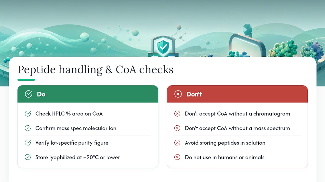
A Certificate of Analysis (CoA) from a reputable supplier will report HPLC purity as a percentage of peak area and include a mass spec confirmation of the expected molecular weight. Those two data points together are what researchers rely on to confirm they have the right molecule at the right purity before running any experiment.
Common examples and misconceptions about peptides and proteins
Named examples worth knowing:
- Insulin (~5,800 Da, 51 residues): sits at the boundary and is variously called a small protein or a peptide hormone depending on context. Its two-chain structure, held together by disulfide bonds, gives it more complexity than a typical short peptide.
- Oxytocin (9 residues): a classic signaling peptide, cyclic, and active at nanomolar concentrations.
- Defensins (18–45 residues): antimicrobial peptides that punch holes in bacterial membranes.
- Collagen: a structural protein built from repeating tripeptide sequences (Gly-X-Y) that assemble into a triple helix, then into fibrils. No short peptide can replicate that architecture.
- Hemoglobin: a quaternary protein with four subunits, each carrying a heme group. Its cooperative oxygen binding is a direct product of that multi-subunit assembly.
Terminology clarifications:
- Polypeptide refers to any chain of amino acids, regardless of length, and is often used for chains in the 50–100 residue range that have not been confirmed to fold stably.
- Peptide fragment describes a portion of a larger protein, often generated by protease digestion for analytical purposes.
- Peptide hormone is a functional label, not a size label; some peptide hormones (like parathyroid hormone at 84 residues) fall in the gray zone.
Common misconceptions:
- Marketing materials frequently use “peptide” to describe any bioactive ingredient, regardless of actual chain length or biological mechanism. That usage is looser than biochemical convention.
- Size cutoffs are conventions. A molecule at 55 residues is not chemically different from one at 45 residues; the label depends on context and the folding behavior of that specific sequence.
- “Protein” does not automatically mean more potent or more complex than “peptide.” Oxytocin, at nine residues, triggers profound physiological responses.
When should you call a chain a peptide versus a protein?
Practical rules for correct usage:
- Fewer than ~50 amino acids: — Default to “peptide” in most contexts. This covers the majority of signaling molecules, synthetic research compounds, and short hormones.
Pro Tip: Before labeling a supplier’s material as a peptide or protein in your lab report, check the CoA for the reported molecular weight and cross-reference it against the sequence. A molecule listed as a “peptide” with a molecular weight above 10,000 Da warrants a closer look at the sequence and folding data.
Casual conversation tolerates more flexibility. In a formal report or publication, precise wording signals scientific literacy and prevents ambiguity in methods sections where the distinction can affect how readers interpret your results.
Why peptide purity and analytical verification matter for research
Structural classification aside, researchers working with peptides face a practical challenge that does not apply in the same way to proteins produced by recombinant expression: chemical synthesis introduces impurities at every coupling step. Achieving 98%+ purity by HPLC requires careful optimization of synthesis conditions and rigorous purification, typically by reversed-phase HPLC.
A reliable CoA for a research-grade peptide should include at minimum: the HPLC chromatogram with retention time and percentage peak area, a mass spectrometry result confirming the expected molecular weight, and the lot-specific purity figure. Neolabpeptides provides third-party-verified CoAs with every product, with purity confirmed at 98%+ by HPLC and mass spectrometry, so researchers can verify identity and purity before any experiment begins. For a detailed walkthrough of what each CoA field means, the guide to reading a peptide Certificate of Analysis covers HPLC and MS readouts step by step.
Storage also affects data quality. Lyophilized peptides should be stored at -20°C or lower, away from moisture and light. Reconstitution in an appropriate solvent immediately before use minimizes degradation. Peptides in solution degrade faster than the lyophilized powder, so preparing only what you need for a single experiment is standard practice.
Pro Tip: When evaluating a peptide supplier’s CoA, look for the HPLC % area figure (not just a stated purity percentage), the mass spec molecular ion confirmation, and the lot number. A CoA without a chromatogram or a mass spectrum is not analytically verified, regardless of what the label says.
All research peptides are for laboratory use only and are not approved for human or veterinary use. Researchers should follow their institution’s protocols for handling, storage, and disposal of research chemicals.
For guidance on peptide purity standards and what to expect from a well-documented research-grade material, Neolabpeptides maintains a detailed lab guide covering interpretation of purity data and proper storage procedures.
Key Takeaways
Peptides and proteins differ primarily in chain length and structural complexity, with the conventional boundary near 50 amino acids, but the functional and analytical consequences of that difference are what matter most in research.
| Point | Details |
|---|---|
| Size-based shorthand | Chains of 2–50 residues are called peptides; chains of 50 or more residues are proteins, though some researchers extend the cutoff up to 100 residues for proteins and describe 50–100 residues as the polypeptide gray zone. |
| Structure drives function | Proteins fold into stable domains enabling enzymes and scaffolds; peptides act mainly as signals and modulators. |
| Stability trade-off | Short peptides are rapidly cleared by proteases and kidneys; strategies like cyclization and D-amino acid substitution extend their half-life. |
| Analytical confirmation | Mass spectrometry and HPLC are the primary tools for confirming molecular identity and purity at the peptide-protein boundary. |
| CoA verification | A valid CoA must include an HPLC chromatogram with % area and a mass spec molecular weight confirmation for the result to be analytically meaningful. |
Why the peptide-protein distinction still matters
The line between peptides and proteins can seem like a technicality, but it carries real consequences for how researchers design experiments, interpret results, and develop therapeutics. A molecule’s size determines how it is synthesized, how quickly it degrades in a biological system, how it is formulated for delivery, and which analytical methods are appropriate for characterizing it. Treating a 60-residue polypeptide as equivalent to a 9-residue signaling peptide in a stability assay, for example, will produce data that cannot be compared across studies.
The terminology also shapes how findings are communicated. When a paper describes a “peptide inhibitor” without specifying residue count or molecular weight, readers cannot assess whether the stability, delivery, and dosing considerations discussed in the methods section are appropriate. Precision here is not pedantry. It is the foundation of reproducible science.
Useful sources for further reading
- Explainer: Peptides vs proteins — what’s the difference? - Institute for Molecular Bioscience - University of Queensland
- Proteins: structure and function | NCBI Bookshelf
- Peptide | Genetics Glossary | genome.gov