

5% Purity Loss in 7 Days? Peptide Stability Rules for Bench Labs
Keep research peptides lyophilized and cold, and treat any reconstituted solution as short-lived. In practice: store lyophilized stock at -20°C or -80°C with moisture excluded, reconstitute into single-use aliquots at a slightly acidic pH, and avoid repeated freeze-thaw cycles. For peptides intended for extended in vivo work, storage discipline alone will not save an unstable sequence — you need structural modification.
TL;DR:
- Store lyophilized peptides at -20°C or -80°C in moisture-free conditions to maximize stability, and keep reconstituted solutions as single-use aliquots at slightly acidic pH.
- Deamidation is most likely with Asn-Gly and Asp-Gly motifs, while oxidation targets methionine, cysteine, and tryptophan; sequence motifs can predict stability risks.
- Repeated freeze-thaw cycles and exposure to light significantly reduce peptide integrity, so aliquot immediately and thaw quickly to limit degradation.
- Buffer choices between pH 4 and 6, along with antioxidants and chelators, can prevent oxidation and hydrolysis, especially when working with sensitive sequences.
- For extended in vivo stability, consider structural modifications like D-amino acids, cyclization, or PEGylation to resist proteolytic degradation and extend circulation time.
Table of Contents
- What Causes Peptide Degradation? Six Chemical Pathways
- Which Residues in Your Sequence Are Stability Risks?
- How Should You Store and Reconstitute Lyophilized Peptides?
- Which Buffers and Excipients Actually Improve Stability?
- Which Modifications Extend a Peptide’s In Vivo Stability?
- How Do You Test Whether a Peptide Batch Is Still Stable?
- What Do Common Degradation Signatures Mean, and When Should You Discard a Batch?
- Neo Lab Peptides: Documentation That Matches Stability Best Practices
- What Actually Moves the Needle in a Working Lab?
- Where to Source Peptides That Hold Up to These Standards
- Where This Article’s Evidence Comes From
- Sources
What Causes Peptide Degradation? Six Chemical Pathways
Peptide stability fails through a small number of well-characterized chemical routes, and most degradation events researchers see on an HPLC trace back to one of six mechanisms. Knowing which pathway threatens a given sequence tells you whether the fix belongs in your buffer, your freezer, or your synthesis order.
Hydrolysis breaks the peptide backbone through nucleophilic attack on the amide bond, and it accelerates in both strongly acidic and strongly basic conditions. Water is the reactant, which is exactly why lyophilized peptides massively outperform peptides sitting in aqueous solution — remove the water and you remove most of the driving force for this pathway.
Deamidation converts asparagine and glutamine side chains into aspartate or glutamate, and it is one of the most reliably observed peptide degradation pathways in room-temperature storage. The reaction runs through a cyclic succinimide intermediate, and it happens fastest when an Asn or Gln sits next to glycine, since glycine’s small side chain lets the backbone fold into the geometry the reaction needs. An Asn-Gly motif in your sequence is a genuine red flag worth checking before you finalize a synthesis order.
Oxidation targets methionine and cysteine almost exclusively, along with tryptophan under stronger oxidative stress. Methionine oxidation to methionine sulfoxide adds a signature +16 Da mass shift that shows up cleanly on LC-MS, and it is driven by dissolved oxygen, trace transition metals, and light exposure working together rather than any single factor. Cysteine’s exposed thiol is even more reactive and can cross-react with other thiols in the same tube.
Diketopiperazine (DKP) formation is a cyclization reaction specific to the N-terminus, where the second residue’s amine attacks the first peptide bond and clips off a cyclic dipeptide fragment. It matters most for dipeptides and short N-terminal sequences, particularly when a proline sits in position two, because proline’s ring geometry favors the intramolecular attack.
Racemization flips an L-amino acid to its D-enantiomer, usually through the same enolizable-proton chemistry that drives some hydrolysis reactions, and it is most common at residues with acidic alpha-protons like aspartate and histidine. A racemized residue does not always destroy activity, but it changes the peptide’s behavior in a receptor-binding assay in ways that are easy to misattribute to a different cause.
Disulfide exchange rearranges cystine bonds in multi-cysteine peptides, scrambling the intended pairing into mispaired isomers, especially above pH 8 where thiol anions become better nucleophiles.
Sequence context routinely predicts trouble before an assay ever runs:
- Asn-Gly and Asp-Gly motifs accelerate deamidation.
- Asp-Pro bonds are unusually acid-labile and prone to hydrolytic cleavage.
- N-terminal Gln cyclizes to pyroglutamate under mildly acidic conditions, a related but distinct N-terminal liability from DKP.
- Any Met or Cys residue is a standing oxidation risk regardless of position.
- Multiple Trp residues raise photo-oxidation risk under ambient lab lighting.
These degradation pathways are residue- and condition-dependent, which is the whole reason a one-size-fits-all storage protocol undersells what careful researchers can actually achieve.
Which Residues in Your Sequence Are Stability Risks?
Every peptide sequence carries a set of predictable liabilities, and reading them off the sequence before you order synthesis saves both money and a failed experiment three weeks later. Run through this checklist against your target sequence:
- Scan for Asn-Gly and Asp-Gly pairs. These are the highest-probability deamidation hotspots, and if one sits in a region critical for receptor binding, deamidation will likely shift or kill activity, not just show up as an inert HPLC shoulder peak.
- Flag every Asp-Pro bond. This motif hydrolyzes disproportionately fast relative to other peptide bonds, especially under mildly acidic reconstitution buffers, and it is worth confirming the bond isn’t sitting in your active pharmacophore.
- Count Met and Cys residues. Each one is an oxidation liability. A single internal methionine is manageable with antioxidant handling; multiple Met or free Cys residues in an unstructured region usually justify a formulation-level fix rather than a redesign.
- Check the N-terminus for Gln, Asn, or Pro in position two. These predict pyroglutamate formation or DKP cyclization, respectively, and both are addressed more reliably by N-terminal acetylation than by storage conditions alone.
- Note any disulfide bonds and their pH exposure window. If your working buffer needs to sit above pH 7.5 for solubility reasons, plan for disulfide scrambling and consider whether a different buffer or a reducing-agent-free workflow is feasible.
Whether a flagged residue calls for re-design or can simply be managed through formulation comes down to where the residue sits functionally. A Met in a flexible linker region tolerates antioxidant-supported storage just fine. A Met inside a binding epitope, where oxidation changes conformation, usually needs replacement with a stability-neutral surrogate like norleucine, or a full re-synthesis with the residue removed.
Common research peptides illustrate the pattern well. BPC-157 contains a Gly-rich backbone that is comparatively hydrolysis-resistant but still benefits from cold, dry storage. IPAMORELIN carries an aromatic-rich sequence where oxidation and light exposure are the dominant concerns, not hydrolysis. CJC-1295, being longer and more structured, is more sensitive to freeze-thaw-induced aggregation than to any single residue-level liability. Checking a candidate sequence against this five-point list takes minutes and routinely prevents a stability failure that would otherwise surface only after a failed bioassay.
How Should You Store and Reconstitute Lyophilized Peptides?
Storage protocol is where most peptide stability is won or lost, and the rules are consistent enough across research peptides to standardize as a lab SOP.
Lyophilized stock should go into a -20°C freezer for working supply and -80°C for long-term archival stock, ideally in a manual-defrost unit to avoid the temperature cycling that frost-free freezers introduce every defrost cycle. Moisture control matters as much as temperature: lyophilized peptides stored cold and dry are substantially more stable than the same peptide in solution, but that advantage disappears if the vial is opened repeatedly at room temperature and allowed to absorb ambient humidity. Desiccant-packed storage, or storage under argon or nitrogen headspace for particularly oxidation-prone sequences, extends shelf life measurably versus a vial left under ordinary lab air.
Reconstitution deserves more care than most protocols give it. Dissolve into a mildly acidic buffer, generally in the pH 4 to 6 range, since most peptides show a preferred stability window between pH 4 and 7, with acid-catalyzed hydrolysis picking up below pH 3 and base-catalyzed hydrolysis and disulfide scrambling picking up above pH 8. Bacteriostatic water is the standard reconstitution solvent for most research peptides; add it slowly down the vial wall rather than directly onto the lyophilized cake, which reduces localized denaturation and foaming. Once reconstituted, split the solution into single-use aliquots immediately rather than drawing repeatedly from one working vial.
Freeze-thaw cycling deserves its own line item because it is the single most common self-inflicted stability failure in a working lab. Each freeze-thaw event stresses the peptide through ice-crystal formation and localized concentration effects at the freezing front, and handling technique measurably affects recovery: slow freezing paired with fast thawing tends to preserve activity better than fast freezing paired with slow thawing in comparable studies. The practical translation is straightforward:
- Aliquot immediately after reconstitution, sized to a single day’s or single experiment’s use.
- Thaw aliquots quickly, at room temperature or in a room-temperature water bath, rather than leaving them on the bench for a slow ambient thaw.
- Never refreeze a thawed aliquot, even if some volume remains unused.
- Label every aliquot with reconstitution date, buffer, and concentration, not just the peptide name.
Shipping and receiving introduce their own risk window. Peptides shipped without cold-chain packaging or with clear vials exposed to light can lose measurable activity before they ever reach your bench, so specify insulated, light-protected packaging when procuring from any supplier and inspect shipments for signs of thermal excursion on arrival.
Pro Tip: Keep a printed reconstitution log taped inside the freezer door listing peptide, date, buffer pH, and aliquot count. It takes ten seconds per entry and it is the single easiest way to catch a forgotten refreeze before it corrupts a whole experiment.
Realistic working-solution lifespans run from a few days at 4°C for oxidation-sensitive sequences up to two to three weeks for more robust peptides, but lyophilized stock at -20°C reliably holds for many months to a year, and archival stock at -80°C longer still. For step-by-step handling protocols, Neolabpeptides’ storage guide walks through timelines by peptide class, and the lyophilization process guide covers cryoprotectant selection for labs freeze-drying their own stock.
Which Buffers and Excipients Actually Improve Stability?
Formulation choices give you a second line of defense against the same degradation pathways storage protocol addresses, and the right buffer or excipient can turn a marginal sequence into a workable one.
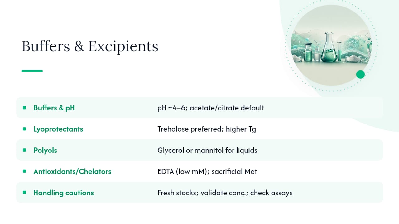
Buffer and pH selection is the foundation. A pH window of roughly 4 to 6 minimizes both hydrolysis and deamidation for most sequences, and acetate or citrate buffers in that range are common defaults for research peptide reconstitution. Push above pH 7.5 only when solubility genuinely demands it, and recognize that doing so raises deamidation and disulfide-scrambling risk in exchange for that solubility gain.
Lyoprotectants matter specifically at the freeze-drying step, since lyophilization itself introduces mechanical and interfacial stress that can partially unfold a peptide even as it removes water. Trehalose is generally preferred over sucrose for longer-term stability since it has a higher glass-transition temperature and does not hydrolyze into reducing sugars the way sucrose can under trace acidic conditions.
Polyols such as glycerol or mannitol serve a related but distinct role in liquid formulations, stabilizing tertiary structure through preferential exclusion from the peptide surface. They are most useful when a peptide must stay in solution rather than lyophilized form for an extended period.
They earn their place primarily in peptides prone to surface-induced aggregation rather than as a universal additive.
Antioxidants and metal chelators directly target the oxidation pathway. Methionine and cysteine oxidation is driven substantially by trace transition metals catalyzing the reaction, so adding a chelator like EDTA at low millimolar concentration binds those metals before they can act. Sacrificial methionine, added free in solution, competes with the peptide’s own methionine residues for oxidative attack and can meaningfully protect the peptide of interest. Inert-gas blanketing with argon or nitrogen during handling and storage reduces dissolved oxygen directly and pairs well with either strategy.
A few contamination cautions apply across all of these. Excipient stocks should be prepared fresh or stored appropriately, since a contaminated sucrose or trehalose stock introduces its own degradation risk. Chelators and antioxidants should be added at the concentrations validated in the literature rather than scaled up on the assumption that more is better. And any excipient introduced into a formulation intended for downstream bioassay work needs to be checked against that assay for interference before you trust the results.
Which Modifications Extend a Peptide’s In Vivo Stability?
Storage and formulation controls protect a peptide sitting in a freezer or a buffer. They do almost nothing against the proteases circulating in serum or tissue, which is why extended in vivo stability generally demands a change to the peptide itself.
D-amino acid substitution replaces one or more L-residues with their D-enantiomer, most often at a protease cleavage site. Proteases are stereospecific for L-amino acids, so a D-substitution at the right position can block cleavage almost entirely. The desmopressin case is the textbook example: substituting a single D-arginine into the vasopressin sequence produced a dramatic increase in serum half-life compared to the natural peptide, turning an impractically short-lived hormone analog into a clinically usable drug.
Cyclization locks the peptide backbone into a constrained ring, which both blocks exopeptidase attack from the termini and often improves binding affinity by pre-organizing the peptide into its bioactive conformation. Head-to-tail cyclization and side-chain cyclization (via lactam or disulfide bridges) are the two dominant approaches, and both trade some synthesis complexity for a real gain in protease resistance.
PEGylation attaches polyethylene glycol chains to the peptide, and it works through steric shielding rather than through blocking a specific catalytic site. The trade-off is genuine: PEGylation reliably extends circulating half-life and reduces renal clearance, but it also often reduces receptor-binding affinity and can complicate downstream purification and characterization.
Backbone N-methylation and peptide stapling target the secondary structure directly. N-methylation of key amide bonds blocks protease recognition motifs while often preserving binding geometry. Stapling, typically via a hydrocarbon bridge between two non-natural residues, locks a helical conformation and simultaneously improves both protease resistance and cell permeability for peptides that need to reach an intracellular target.
Choosing among these options is a matter of what’s failing and what you can afford to lose. If proteolysis at a specific site is the dominant failure mode, D-substitution or N-methylation at that site is the more surgical fix. If the whole peptide is short-lived because of general exopeptidase and renal clearance, cyclization or PEGylation addresses the broader problem, at some cost to binding affinity or synthesis simplicity. Sequence-level anti-degradation strategies are rarely interchangeable substitutes for one another; they solve different failure modes, and a peptide with both a protease-cleavage problem and a general clearance problem often benefits from combining two approaches.
When protease resistance alone still leaves clearance too fast, pairing a modified peptide with a delivery vehicle, lipid nanoparticles or exosome-based carriers being the most actively studied options, extends circulation time further by shielding the peptide from filtration and enzymatic exposure altogether. This is generally a later-stage consideration once bench-level modification has already been optimized, not a first-line fix for a peptide still in early characterization.
How Do You Test Whether a Peptide Batch Is Still Stable?
Determining whether a batch has degraded past usability requires a defined assay workflow, not a visual check of the vial.
- Run RP-HPLC as the first-line stability-indicating assay. A shift in retention time, a new shoulder peak, or peak broadening relative to the reference chromatogram flags degradation before you need mass spec to characterize it. Stability-indicating RP-HPLC and LC-MS remain the standard tools for this purpose across the field.
- Follow up ambiguous peaks with LC-MS. A +16 Da mass shift on the intact peptide or a tryptic fragment points to oxidation, almost always at Met or Cys. A shift consistent with a loss or gain of roughly 1 Da, paired with a change in HPLC retention time for an otherwise identical mass, points to deamidation via the succinimide intermediate.
- Set a numeric acceptance threshold before you need one, not after. A widely used rule of thumb treats greater than 5% purity loss over 7 days at 37°C as a red flag warranting investigation, and applying that threshold consistently keeps QC decisions from becoming subjective judgment calls made batch by batch.
- Use serum half-life and proteolysis assays for anything destined for in vivo or cell-based work. Spiking the peptide into serum or a protease cocktail and sampling over time by HPLC or LC-MS gives you a functional half-life estimate that a static purity assay cannot, since it captures enzymatic cleavage that bench storage conditions never test for.
- Set a sampling cadence proportional to how the peptide will be used. A peptide going straight into a same-week assay needs a single confirmatory HPLC run. A peptide intended for a multi-month study justifies periodic re-testing, for instance at baseline, one month, and three months, especially if it is stored in solution rather than lyophilized.
A single HPLC purity number at time zero tells you almost nothing about whether a peptide will still be intact when you actually run your assay three weeks later. The number that matters is the slope of purity loss over time under your actual storage conditions, not the number on the Certificate of Analysis alone.
Interpreting a borderline result matters as much as generating it. A peak that grows measurably between two time points sampled under the same storage condition is the signature you should actually act on.
What Do Common Degradation Signatures Mean, and When Should You Discard a Batch?
Reading an HPLC or LC-MS result correctly is what separates a fixable formulation problem from a batch that belongs in the sharps container.
A new peak eluting earlier than the main peak, paired with a mass shift consistent with hydrolysis fragments, usually points to backbone cleavage, most often at an Asp-Pro bond or under storage conditions that drifted outside the pH 4 to 7 window. A +16 Da shift on LC-MS with no retention-time change in the parent peak signature is close to diagnostic for methionine oxidation. A shoulder peak with an isobaric or near-isobaric mass to the main peak usually indicates deamidation, isomerization, or racemization, distinguishable mainly by which residue position the peptide’s known liabilities point to. Broadened or split peaks with no clear mass shift often indicate aggregation rather than covalent degradation, and that distinction matters because aggregation sometimes reverses with a buffer change while covalent degradation does not.
Immediate mitigation depends on which signature you’re looking at:
- For oxidation signatures, add a metal chelator and switch to inert-gas storage going forward; this won’t reverse damage already done but will slow further loss in the remaining stock.
- For hydrolysis or deamidation trending upward across sampling points, move the working stock to a lower storage temperature and recheck the reconstitution buffer’s pH.
- For aggregation, try a mild surfactant addition or a brief buffer exchange before assuming the batch is lost.
Longer-term fixes belong at the sequence level: if a particular residue reliably degrades across multiple batches regardless of storage discipline, re-synthesis with a stability-oriented substitution, D-amino acid swap, N-methylation, or removal of the offending motif, is the more permanent answer than repeated remediation.
The decision rule is simple in practice. If purity loss exceeds threshold, the degradation product is unidentified, or the affected region overlaps your peptide’s active site, discard and resynthesize rather than trying to rescue the batch. Recognizing oxidation-specific signatures early is usually cheaper than repeating a failed downstream experiment.
Neo Lab Peptides: Documentation That Matches Stability Best Practices
That documentation matters for the exact reasons this article covers: a COA gives you the baseline purity number your own stability-indicating assays get compared against later, and lot-specific mass spec data helps you confirm you’re starting from an unoxidized, undegraded stock before you ever open the vial.
Products ship in lyophilized form, the storage state this article treats as the foundation of peptide stability, specifically to give researchers control over reconstitution timing, buffer selection, and aliquoting rather than receiving a peptide that has already spent unknown time in solution. Handling instructions accompany each order to support correct freezer temperature and moisture-control practices from the moment a shipment arrives.
For implementation detail beyond what a COA alone conveys, Neolabpeptides’ third-party testing guide explains what the verification process actually checks, and the lyophilized peptide handling guide walks through storage and reconstitution SOPs matched to the practices described above.
What Actually Moves the Needle in a Working Lab?
Most stability failures in a working lab trace back to two or three preventable habits, not to some exotic degradation chemistry nobody saw coming. Cold, dry, lyophilized storage with single-use aliquoting gets you most of the available stability gain for almost no added cost or complexity. Everything past that, buffer optimization, excipient tuning, sequence modification, delivers real but progressively smaller returns for progressively more effort.
Labeling discipline is the most underrated control in this entire discussion. An unlabeled or vaguely labeled aliquot gets left on a bench, refrozen by mistake, or grabbed for the wrong experiment, and none of that shows up until a result doesn’t replicate. Date every aliquot, note the reconstitution buffer, and store working stock separately from archival stock so nobody warms the wrong vial reaching for the one they actually need.
Bringing in a contract lab for formal stability testing makes sense once a peptide moves from a single experiment toward a program, a multi-month study, a formulation destined for further development, where the cost of a formal accelerated-stability study is small next to the cost of a compromised dataset. For routine bench work, disciplined storage and periodic HPLC checks cover the risk adequately.
— Stephan
Where to Source Peptides That Hold Up to These Standards
Every control this article covers, cold lyophilized storage, verified purity, documented handling, only matters if the peptide arrived stable in the first place.

The catalog covers the research peptides labs actually reach for, IPAMORELIN, CJC-1295, TB500, BPC-157, GLP-1 analogs, NAD+, GHK-Cu, along with bacteriostatic water for reconstitution, all shipped lyophilized with fast delivery across the U.S. and handling documentation included so your storage protocol starts correctly on day one. Every product ships for laboratory research use only, not for human or veterinary use. Check the Certificate of Analysis and storage instructions for any peptide before you order by visiting the Neolabpeptides product catalog and confirming the lot documentation matches the purity and handling standards this guide describes.
Where This Article’s Evidence Comes From
The degradation mechanisms and modification strategies covered here draw substantially on a peer-reviewed review of in vivo degradation forms and anti-degradation strategies for therapeutic peptides and a companion review of strategies for improving peptide stability and delivery, both published on PubMed Central.
Storage and formulation guidance reflects supplier-level technical documentation from Sigma-Aldrich’s peptide stability technical article and a broader formulation overview from ScienceDirect’s peptide stability topic summary. Assay design and QC threshold guidance references a technical guide to peptide degradation pathways and stability prevention.
Sources
- In vivo degradation forms, anti-degradation strategies, and clinical applications of therapeutic peptides in non-infectious chronic diseases - PMC
- Peptide Stability and Potential Degradation Pathways (Sigma-Aldrich technical article)
- Peptide Stability - ScienceDirect Topics