

Avoid Dosing‑Day Failures: GLP‑1 Checklist for Preclinical Studies
In this context, “GLP” refers to GLP‑1 research peptides, not Good Laboratory Practice. The priority action before any study begins is confirming reagent identity and purity through HPLC/MS Certificates of Analysis, then designing PK sampling windows around the compound’s actual half‑life. Long‑acting analogs can run 24 to over 60 hours in preclinical species, and fatty‑acid modifications change how the molecule behaves in your assay, not just in the animal.
TL;DR:
- Confirm reagent purity and identity with third-party HPLC and mass spectrometry before starting any study to avoid incorrect bioanalytical results.
- Design PK sampling windows based on the actual half-life of the analog, which can vary from 24 to over 60 hours depending on modifications.
- Pair each PK time point with a matched PD measurement, such as glucose tolerance or body weight, to accurately correlate exposure and effect.
- Use multiple sampling points at both Cmax and trough to detect exposure-dependent effects and account for accumulation over repeated doses.
- Verify peptide stability during storage by running short-term stability tests linked to the certificate of analysis to prevent variability in long-term studies.
Table of Contents
- What Are GLP‑1 Peptides and Analogs Used in Preclinical Research?
- How Should PK/PD Study Design Account for Species and Timing?
- What Bioanalytical Challenges Come With Quantifying GLP‑1 Peptides?
- What Purity and Analytical Standards Should GLP‑1 Reagents Meet?
- How Should Teams Handle Formulation, Storage, and Dosing?
- What Does a Practical GLP‑1 Preclinical Checklist Look Like?
- Neo Lab Peptides: A Documented Source for Research‑Grade GLP‑1 Reagents
- The Gap Most Teams Miss Before Dosing Day One
- Sources
What Are GLP‑1 Peptides and Analogs Used in Preclinical Research?
Native GLP‑1 fragments degrade within minutes, which is why almost every reagent used in preclinical pharmacology today is a modified analog built for a longer working window. Structural tweaks like Aib or Ala substitutions at the DPP‑4 cleavage site resist enzymatic breakdown, while lipidation, attaching a fatty‑acid chain that binds albumin, extends circulating time dramatically. That albumin binding is a double‑edged design feature. It stretches half‑life, but it also means the peptide travels bound to a carrier protein in plasma, which affects how you extract and detect it later in the bioanalytical lab.
Beyond single‑target analogs, co‑agonists that hit GLP‑1 and glucagon receptors together, or triple agonists layering in GIP, are increasingly common in efficacy pipelines. Receptor balance between these targets does not behave identically across species, so a construct that looks clean in one model can produce a species‑dependent effect profile in another. Know which construct you’re running before you design anything downstream.
How Should PK/PD Study Design Account for Species and Timing?
Terminal half‑life and Tmax dictate almost everything else in your study design. Sample too early and you miss the exposure peak; sample too late and you’re reading noise instead of signal. One long‑acting GLP‑1 analog reported a terminal half‑life of 61.3 hours with a Tmax around 14 hours in cynomolgus monkeys, which is a completely different sampling problem than a native fragment cleared in single‑digit minutes. Plan your blood draws around the actual molecule, not a generic peptide template.
Rodent and nonhuman primate models rarely agree on magnitude, and sometimes not even on direction. A protracted GLP‑1/glucagon co‑agonist produced dose‑dependent weight loss in mice, but the split between lean and fat mass loss varied by species, and glucose tolerance outcomes depended heavily on how long the study ran and what exposure level the animals actually reached.
Design choices that reduce translational risk include:
- Sampling at both Cmax and trough (Cmin), not just one timepoint, to catch exposure‑dependent effects that a single draw would miss.
- Running glucose tolerance tests at multiple exposure windows rather than assuming an acute response predicts steady‑state control.
- Tracking accumulation across repeated dosing, since long half‑lives mean day‑10 trough levels can look nothing like day‑1 trough levels.
- Pairing every PK timepoint with a matched PD readout: body weight, food intake, insulin, or glucose tolerance, so exposure and effect can actually be correlated.
Functional signaling adds another layer worth building into your endpoints. β‑arrestin recruitment versus cAMP signaling can diverge even when receptor binding looks identical on paper, and biased agonism has been shown to alter acute versus sustained GLP‑1R responses. If your efficacy readout only captures binding affinity, you may be missing the mechanism that actually drives the chronic result.
What Bioanalytical Challenges Come With Quantifying GLP‑1 Peptides?
Peptide bioanalysis fails most often at the assay design stage, not the instrument. GLP‑1 analogs circulate at low plasma concentrations and often bind albumin, so LC‑MS/MS methods need specific MRM transitions and dedicated sample enrichment to detect the compound reliably.
Practical fixes that matter more than people expect:
- Screen CID spectra and select higher m/z fragment ions for MRM transitions rather than defaulting to the first fragment that shows a signal.
- For fatty‑acid–modified peptides, add a dissociation step before SPE to release the peptide from albumin before extraction, or recovery numbers will look artificially low.
- If carryover or peptide “stickiness” shows up between injections, try alternative column chemistries like HILIC and rework the needle wash program before blaming the compound.
- Validate recovery across the full concentration range you expect in‑study, not just at the midpoint, and check matrix effects against blank plasma from the actual species you’re dosing.
Pro Tip: Run a quick dissociation‑step comparison (with versus without) on your first pilot samples before committing to a full validation. Albumin‑bound peptides can show two‑fold or greater recovery differences depending on whether that step is included, and finding out mid‑study is expensive.
Long‑acting peptides with complex elimination profiles also benefit from radiolabeled ADME work and extended mass balance sampling to fully characterize metabolites rather than relying on a standard short sampling window.
What Purity and Analytical Standards Should GLP‑1 Reagents Meet?
A peptide’s Certificate of Analysis is only as good as the methods behind it, and single‑method verification is not enough for reagents going into a preclinical dataset. Orthogonal analytics meaning RP‑HPLC or UPLC for purity, LC‑MS for identity and molecular weight, and SEC for aggregation, catch different failure modes that a single test would miss entirely.
Common impurities worth screening for specifically:
- Truncated sequences from incomplete synthesis, which can retain partial receptor activity and confound dose‑response data.
- Oxidized or deamidated variants that shift charge and mass just enough to alter receptor binding without showing up on a casual visual inspection.
- Aggregates detectable by SEC that a standard RP‑HPLC purity run would completely miss.
- Residual host‑cell or synthesis‑related impurities that can introduce unwanted background activity in functional assays.
A robust CoA documents purity by HPLC, confirms identity by mass spec, states the lot number, and ideally comes from third‑party verification rather than in‑house testing alone. Even small structural drift in a batch can shift receptor binding enough to muddy an efficacy result, so treat analytical files as part of the study record, not paperwork to file away after the order arrives.
How Should Teams Handle Formulation, Storage, and Dosing?
Reconstitute lyophilized peptide with the solvent the CoA specifies, and add it slowly along the vial wall rather than directly onto the powder to limit aggregation. Aliquot into single‑use, low‑protein‑binding tubes at your working concentration immediately after reconstitution rather than refreezing a stock vial repeatedly.
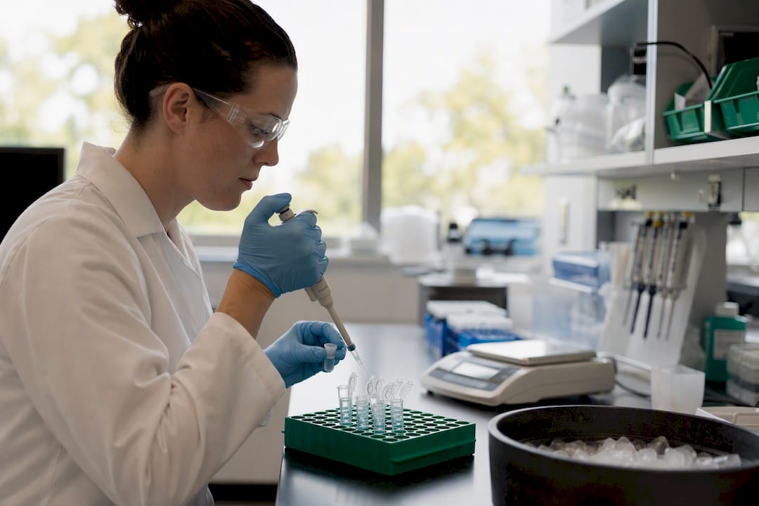
For subcutaneous dosing, formulation stability at the injection concentration matters more than most protocols acknowledge. If you’re testing a noninjectable route, permeation enhancers like sodium caprate can markedly improve bioavailability for GLP‑1 analogs crossing intestinal membranes, but that only helps if the peptide is intact when it gets there.
Before committing to a chronic study, run a short bench stability check: keep a QC aliquot under study conditions for the expected dosing period and test it against a fresh reference. Link every study vial to its CoA and lot number, and keep those batch records with the study file, not separately.
What Does a Practical GLP‑1 Preclinical Checklist Look Like?
- Pre‑study: Verify the CoA against third‑party HPLC/MS results, run a quick solubility and short‑term stability check, and map out PK sampling windows based on the analog’s expected half‑life.
- In‑study: Collect PK samples at both Cmax and trough, pair every timepoint with a relevant PD biomarker such as glucose tolerance or body weight, and track food intake alongside dosing.
- Post‑study: Archive retained plasma samples, run metabolite identification if the PD result doesn’t match expectations, and preserve all analytical documentation with the final study file.
Neo Lab Peptides: A Documented Source for Research‑Grade GLP‑1 Reagents
Every checklist item above depends on one thing: trusting what’s in the vial. Neo Lab Peptides supplies research‑grade GLP‑1 analogs verified at over 98% purity through third‑party HPLC and mass spectrometry, with a Certificate of Analysis included on every order.

That documentation does real work before your study even starts. Instead of running your own identity confirmation from scratch, you can move straight into PK planning and assay setup, because the purity and identity data arrive with the shipment rather than after a separate round of internal testing. Products like the GLP1‑S semaglutide‑style research peptide and the GLP3‑RT triple agonist reagent ship lyophilized with lot‑specific CoAs attached, and all products are labeled for laboratory research use only, not for human or veterinary administration.
If your team is scoping a new efficacy or mechanistic study, visit Neo Lab Peptides to review current CoAs and request a sample kit before your next study timeline locks in.
The Gap Most Teams Miss Before Dosing Day One
The most common failure in GLP‑1 preclinical work isn’t a bad hypothesis. It’s skipping analytical verification or sampling PK at the wrong exposure window, then trying to explain an inconsistent PD result after the fact. Both problems get caught early with the same two habits: confirm the CoA against independent HPLC/MS data before the compound touches an animal, and build in PK bridging timepoints at both peak and trough exposure. Neither takes much extra time. Both save a study.
— Stephan
Sources
- Journal article on permeation enhancers improving oral/permeation bioavailability (2025)
- Pharmacokinetic profiling of long‑acting GLP‑1 analogs in nonhuman primates (example data)
- Analysis and characterization of GLP‑1 peptides (The Analytical Scientist, 2025)
- Preclinical evaluation of a protracted GLP‑1/glucagon receptor co‑agonist: Translational difficulties and pitfalls (PLOS ONE)